Need help? Call today!
+1-617-398-7852
Microscopes are indispensable tools in science, medicine, industry, and research. But with so many variants out there, you might ask: What are the 4 types of microscopes? In this blog post, I'll break them down, explain how each works, and highlight where each is most useful. Whether you're a student, lab technician, or science enthusiast, this guide will help you pick the right microscope (or just understand their differences better).
When people ask me, “What are the 4 types of microscopes?”, they’re often referring to the main ways microscopes produce images and the types of samples they’re designed to study. In microscopy, “type” can refer to the imaging technology used (such as light, electrons, or probes) or the practical application (like biological, metallurgical, or live-cell imaging).
At its core, microscopes are classified based on how they interact with the sample to create a magnified image. The four most widely recognized categories are:
Optical (Light) Microscopes – use visible light and glass lenses to magnify cells, tissues, and small organisms.
Electron Microscopes – rely on beams of electrons instead of light to achieve nanometer-scale resolution.
Scanning Probe Microscopes – use a physical probe that “feels” the surface at the atomic level for 3D topographical mapping.
Confocal / Fluorescence Microscopes (Advanced Optical) – employ lasers, fluorescent dyes, and digital scanning to capture high-contrast, 3D images of labeled structures.
Each of these microscope types offers unique advantages depending on what needs to be observed. That depends on whether it’s live biological cells, metal surfaces, nanoparticles, or molecular structures. Understanding their differences helps you choose the right microscope for your laboratory or educational purpose.
It’s important to note that different sources classify microscopes differently. For example, some educational materials (like those from Evolve Scientific or Microscope World) categorize the four main types as compound, stereo, digital, and inverted microscopes. All of which are actually sub-categories of optical microscopes. These are useful for teaching contexts and general lab work, while the broader scientific classification (optical, electron, scanning probe, and confocal) represents the technological basis of modern microscopy.
In the next sections, we’ll explore each of these four main types of microscopes in detail and explain how they work, their key features, applications, advantages, and limitations. That way, you can clearly understand which type best fits your scientific or educational needs.
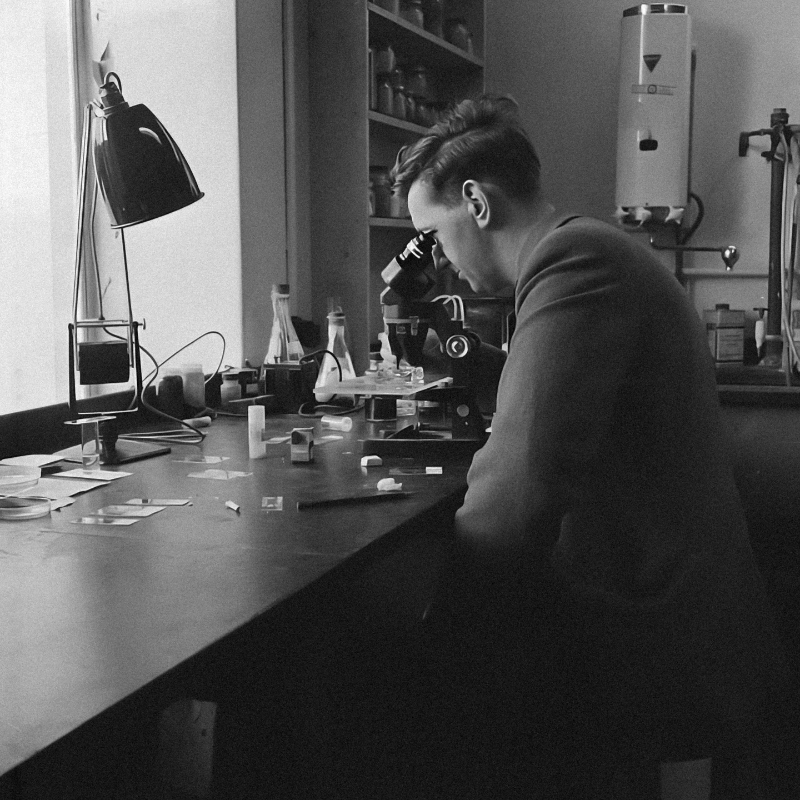
The optical microscope, also known as the light microscope, is the most common and widely used type of microscope. It operates by using visible light and optical lenses to magnify specimens, allowing scientists, educators, and technicians to observe structures that are too small for the naked eye. When people ask me “What are the 4 types of microscopes?”, the optical microscope is typically the first that comes to mind. Which formed the foundation of modern microscopy.
Optical microscopes function by directing light from a built-in lamp or LED through or onto the specimen. This light passes through a series of lenses that bend (refract) and magnify the image.
The objective lenses (closest to the specimen) provide the initial magnification.
The eyepiece lenses (also called ocular lenses) further enlarge the image for viewing.
The magnified image can be seen directly through the eyepiece or captured by a digital camera for display on a screen.
The resolution of optical microscopes is limited by the wavelength of visible light, typically achieving detail down to around 200 nanometers. While this limits their ability to visualize structures smaller than that (like viruses or molecular complexes), optical microscopes remain invaluable for routine laboratory and educational use.
There are several subtypes of optical microscopes, each designed for specific viewing needs or specimen types.
Compound Microscope – Uses multiple lenses to achieve high magnification (up to 1000x). Commonly used in biology labs for observing cells, tissues, and microorganisms.
Stereo or Dissecting Microscope – Offers lower magnification but provides a 3D view of larger specimens like insects, minerals, circuit boards, or fossils.
Phase-Contrast Microscope – Enhances contrast in transparent, unstained specimens, such as living cells, without the need for dyes.
Darkfield Microscope – Illuminates specimens against a dark background, revealing fine details invisible under standard brightfield lighting.
Differential Interference Contrast (DIC) Microscope – Produces high-contrast, pseudo-3D images of live or transparent specimens, ideal for cell biology.
Digital / USB Microscope – Combines optical magnification with a digital camera sensor, displaying images on a monitor for easy sharing or documentation. Frequently used in education, forensics, and electronics.
Each subtype has unique optical configurations that make it ideal for different scientific, industrial, or educational applications.
Optical microscopes are used across a wide range of fields because of their versatility and simplicity:
Biology & Medicine: Examine cells, bacteria, tissues, and microorganisms.
Education: Demonstrate cellular structures and basic microscopy techniques.
Pathology & Diagnostics: Identify disease-causing microorganisms and tissue abnormalities.
Industrial Inspection: Analyze circuit boards, textiles, and manufactured components.
Forensics & Archaeology: Examine fibers, artifacts, and evidence samples.
Because of their balance of ease of use, affordability, and real-time visualization, optical microscopes remain the go-to tool for laboratories, classrooms, and research facilities worldwide.
Strengths:
Simple to operate and maintain
Affordable compared to electron or probe-based microscopes
Allows real-time observation of live samples
Available in portable and digital formats for flexible use
Limitations:
Resolution is limited by light wavelength (~200 nm), so ultrastructural details (like organelles or viruses) are not visible
Requires transparent or thin samples for transmitted-light imaging
Limited magnification compared to electron microscopy
Electron microscopes are powerful instruments that use a focused beam of electrons instead of visible light to form images of specimens. Because electrons have much shorter wavelengths than light, these microscopes achieve ultra-high resolution which can be up to a thousand times greater than the best optical microscopes. When people ask “What are the 4 types of microscopes?”, the electron microscope stands out as the tool that allows us to visualize the nanoscopic world in incredible detail.
Electron microscopes rely on electromagnetic lenses to focus electrons onto a specimen. The interaction between the electrons and the sample produces signals that are captured by detectors and converted into images.
Key operational characteristics include:
Samples must often be placed in a vacuum chamber, since air molecules can interfere with the electron beam.
For Scanning Electron Microscopy (SEM), specimens are typically coated with a thin conductive layer (like gold or carbon) to enhance signal quality.
Transmission Electron Microscopy (TEM) requires ultra-thin specimens so that electrons can pass through the sample.
The resulting images are displayed on a monitor and can reveal features as small as 0.1 nanometers, depending on the type of instrument.
There are two main types of electron microscopes, each designed for distinct imaging needs.
Transmission Electron Microscope (TEM)
Function: Electrons pass through an extremely thin specimen, revealing internal structures in high detail.
Output: Produces a 2D image that shows the internal ultrastructure of cells, organelles, or materials.
Applications: Cell biology, virology, and nanomaterials research
Scanning Electron Microscope (SEM)
Function: Electrons scan across the surface of a specimen, detecting scattered electrons to form a 3D-like surface image.
Output: Provides a detailed view of surface morphology and texture.
Applications: Metallurgy, materials science, forensics, and failure analysis.
Both TEM and SEM have revolutionized fields such as nanotechnology, biomedical imaging, and materials characterization, giving scientists insight into structures far smaller than what optical systems can detect.
Electron microscopes play a vital role across scientific and industrial research:
Biology & Medicine: Imaging viruses, organelles, and subcellular components.
Material Science & Metallurgy: Analyzing crystal structures, metals, and composites.
Nanotechnology: Studying nanoparticles, thin films, and molecular assemblies.
Engineering & Failure Analysis: Examining fractures, corrosion, and defects at the microscopic level.
Forensics & Semiconductor Research: Inspecting residues, microchips, and contamination in manufacturing.
These applications highlight why electron microscopes are essential tools wherever precision and detail are critical.
Pros:
Extremely high resolution - up to sub-nanometer detail.
Ability to visualize ultrastructures beyond light microscopy’s limits.
Produces both surface (SEM) and internal (TEM) information.
Crucial for cutting-edge research in nanoscience and medicine.
Cons:
High cost - both for purchase and maintenance.
Complex sample preparation, often requiring dehydration or coating.
No live imaging - samples must be placed in a vacuum, making the process destructive or non-reversible.
Requires trained operators and controlled environments.
Scanning Probe Microscopes (SPMs) represent one of the most advanced categories of microscopes, capable of imaging surfaces at the nanometer or even atomic scale. Instead of using light or electron beams, SPM technology employs a fine mechanical probe that physically interacts with the sample’s surface. This allows researchers to "feel" and map the surface topography with incredible precision.
When discussing “What are the 4 types of microscopes?”, scanning probe microscopy stands out for its ability to image individual atoms, a level of resolution unmatched by optical or electron microscopes.
The fundamental principle of SPM is simple yet powerful:
A sharp probe tip which is only a few atoms wide is scanned across a sample surface in a highly controlled manner. As the tip moves, it experiences interactions (such as forces or currents) with the atoms on the surface. These interactions are measured and converted into a digital signal, which is then processed to create a 3D image or topographic map of the surface.
SPMs often use piezoelectric scanners, which can move the tip with sub-nanometer precision in the x, y, and z directions. Depending on the type of interaction measured, several specific instruments fall under the SPM category.
Atomic Force Microscope (AFM)
How it works: A tiny cantilever with a sharp tip scans the sample’s surface. As the tip moves, forces between the tip and surface atoms cause the cantilever to deflect.
Detection: A laser beam reflects off the cantilever into a photodiode detector, recording these deflections and translating them into topographical data.
Applications: AFM is used to study polymers, cells, DNA, and nanomaterials. It can measure mechanical properties like stiffness and elasticity.
Scanning Tunneling Microscope (STM)
How it works: Based on the principle of quantum tunneling, STM uses a conductive tip positioned extremely close to a conductive or semiconductive surface.
Detection: When a voltage is applied, electrons tunnel between the tip and the surface, creating a measurable current. Variations in this current reveal the atomic structure of the surface.
Applications: STM was the first instrument to visualize individual atoms directly. It’s widely used in surface science, materials engineering, and nanotechnology research.
Scanning Probe Microscopy has transformed how scientists study materials at the nanoscale. Key applications include:
Nanotechnology: Imaging and manipulating atoms or molecules for nanoscale engineering.
Surface Science: Analyzing roughness, grain boundaries, and surface chemistry.
Material Research: Measuring electrical, magnetic, and mechanical properties at ultra-small scales.
Biological Sciences: Studying molecular structures, cell membranes, and protein folding.
Semiconductors: Evaluating defects and surface uniformity in microelectronic components.
SPMs provide not just visual information but also quantitative data about forces, conductivity, and elasticity, making them essential for both physical and life sciences.
Strengths:
Atomic or near-atomic resolution, far surpassing traditional microscopes.
Can operate in ambient air, liquid, or vacuum environments, depending on configuration.
Provides 3D surface topography and quantitative property measurements.
Enables nanoscale manipulation while some versions can even move individual atoms.
Limitations:
Limited imaging area which typically is only a few micrometers.
Slow scan speed, since the tip must physically trace each line of the surface.
Only captures surface details, not internal structures.
Requires vibration isolation and precise control to maintain accuracy.
Confocal and fluorescence microscopes represent the cutting edge of optical microscopy, bridging traditional light microscopy and modern digital imaging. These advanced systems use lasers, fluorescent dyes, and computational imaging to produce high-contrast, three-dimensional views of cells, tissues, and biological processes in real time.
When answering “What are the 4 types of microscopes?”, this category often referred to as advanced optical microscopy includes the most sophisticated light-based imaging methods used in biomedical research, neuroscience, and cell biology.
These microscopes share a foundation in light excitation and emission, but they achieve imaging in different, complementary ways:
Fluorescence Microscopy:
Uses fluorescent dyes or genetically encoded fluorophores that attach to specific molecules within a specimen.
When illuminated with light of a specific wavelength, these dyes absorb and re-emit light at a longer wavelength.
This emission is captured through optical filters, allowing scientists to view only the labeled structures such as proteins, organelles, or DNA against a dark background.
The result is a high-contrast image that highlights selected cellular components with remarkable clarity.
Confocal Microscopy:
Uses a laser scanning system that focuses light on a very narrow focal plane within the specimen.
A pinhole aperture blocks out-of-focus light, dramatically increasing image sharpness.
By scanning multiple planes at different depths, the microscope can reconstruct 3D images of thick samples or whole cells.
The method is especially powerful for live-cell imaging and 3D tissue visualization.
Together, these approaches enable researchers to observe biological structures and processes in real time, often at the molecular level.
Advanced optical microscopes have become indispensable tools in modern science, particularly in the biological and medical fields.
Cell Biology: Visualizing proteins, organelles, and intracellular interactions.
Neuroscience: Mapping neural connections and observing synaptic activity.
Developmental Biology: Studying embryo development and gene expression.
Pathology: Identifying diseased tissues or cancer markers via fluorescence tagging.
Live-Cell Imaging: Tracking dynamic processes like mitosis or molecular transport in living cells.
Because of their ability to selectively image specific molecules with high contrast, these microscopes have revolutionized molecular diagnostics, genetic engineering, and drug discovery.
Strengths:
Enables specific molecular imaging through targeted fluorescent labeling.
Produces high-contrast and 3D reconstructions of complex biological structures.
Allows for live-cell imaging, depending on sample preparation and equipment.
Excellent for studying protein localization, dynamics, and interactions.
Limitations:
Photobleaching - fluorophores lose brightness over time due to repeated exposure.
Phototoxicity - intense laser light can damage live specimens.
High cost - advanced optics, lasers, and detectors make these systems expensive.
Complex operation - requires specialized training and image-processing expertise.
Despite these challenges, confocal and fluorescence microscopy remain the gold standard for cellular and molecular imaging due to their precision and versatility.
| Type | Imaging Principle | What You Can See | Typical Uses | Pros / Strengths | Limitations |
|---|---|---|---|---|---|
| Optical (Light) Microscopes | Visible light + lenses | Cells, tissues, small organisms, surfaces | Biology labs, education | Easy, real-time, lower cost | Limited resolution; can’t see ultrastructure |
| Electron Microscopes (TEM, SEM) | Electron beams | Fine internal structures (TEM), surfaces (SEM) | Nanotech, cell ultrastructure, materials | Very high resolution, ultrastructure imaging | Expensive, complex prep, vacuum required |
| Scanning Probe Microscopes (SPM) | Mechanical probe scanning | Surface topography at atomic / nanoscale | Nanoscience, surface phenomena | Extreme surface resolution | Only surface imaging; slow; limited area |
| Confocal / Fluorescence Microscopes | Laser scanning / fluorophores | Labeled molecules, 3D structure, live-cell dynamics | Cell biology, 3D imaging, live imaging | Molecular specificity, optical sectioning | Photobleaching, cost, complexity |
The question “What are the 4 types of microscopes?” doesn’t have a single, universally “correct” answer simply because microscopes can be classified in different ways depending on the context. The classification system used often reflects whether the focus is educational, industrial, or scientific research.
Microscopes can be categorized by:
Imaging Method: How the image is formed (optical, electron, probe, or fluorescence).
Sample Type: Whether the microscope examines biological, metallurgical, or materials samples.
Function or Design: Based on physical structure or viewing style (e.g., compound, stereo, inverted, digital).
For instance, educational and teaching materials often define the four types as:
Compound Microscope - for viewing cells and thin biological specimens.
Stereo Microscope - for 3D viewing of larger objects.
Inverted Microscope - for live-cell imaging and culture observation.
Digital Microscope - for computer-based image capture and display.
This set emphasizes practical usage and classroom relevance.
In research and industrial contexts, however, the four main types of microscopes are generally recognized as:
Optical (Light) Microscopes
Electron Microscopes
Scanning Probe Microscopes
Confocal / Fluorescence (Advanced Optical) Microscopes
This classification reflects how microscopy is divided in scientific literature, engineering applications, and biotechnology research. Each type represents a major technological advancement that expanded what scientists can see and measure from micrometer-scale cells to individual atoms.
This modern, research-focused framework is widely adopted because it’s based on fundamental imaging principles rather than instrument design. It distinguishes microscopes by how they generate images through light, electrons, or physical probes and recognizes the role of advanced optical methods like fluorescence and confocal microscopy.
Such a classification helps students, engineers, and researchers understand:
The underlying physics behind each type of microscope.
The scale of resolution achievable (from micrometers to angstroms).
The scientific domains where each technology is most useful (biology, nanotechnology, materials science, etc.).